
Abstract
A 32-year-old male patient presented with uncontrolled convulsions to the emergency room. He had epilepsy since childhood and was on tablet phenytoin sodium 100 mg three times a day. However, on detailed history elicitation and clinical examination, he was found to have a constellation of findings. He had multiple swellings over both the lower limbs and upper limbs at the site of various tendon insertions (xanthomas), mental retardation, speech disturbance, bilateral pyramidal tract involvement and bilateral cataracts. Based on clinical features, a diagnosis of cerebrotendinous xanthomatosis: a relatively rare genetic disorder was suspected, and later on confirmed histopathological.
See this image and copyright information in PMC
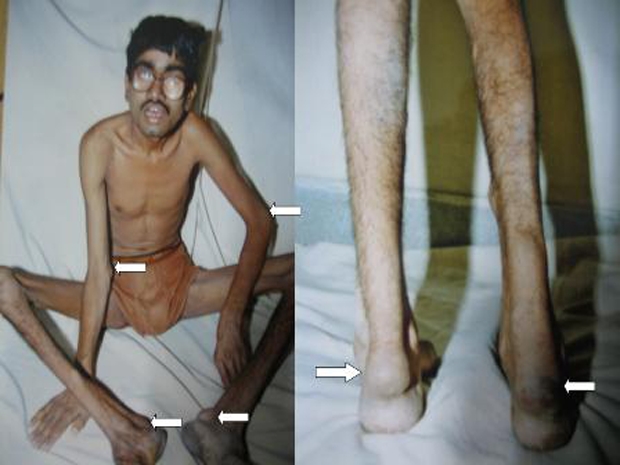
Figure 1 Arrows showing large tendon xanthomas over the Achilles tendon, elbow and triceps tendon. Black arrow indicates the patient is wearing thick glasses due to aphakia.
See this image and copyright information in PMC
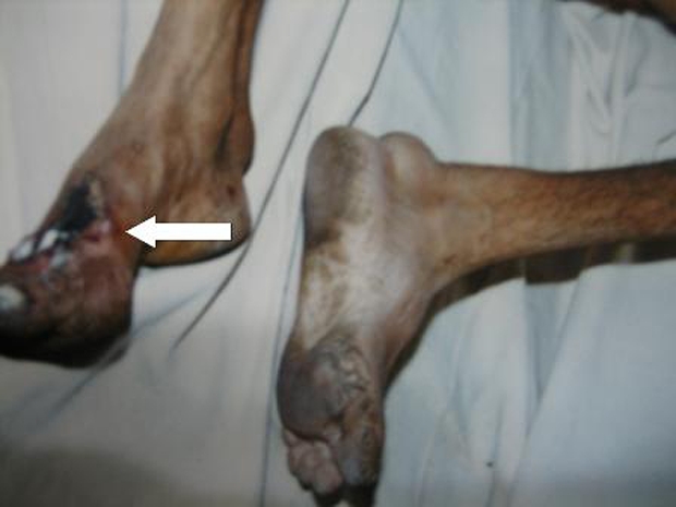
Figure 2 Healed ulcer over the dorsum of left foot.
See this image and copyright information in PMC
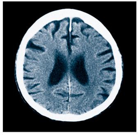
Figure 3 CT of head showing mild cerebral atrophy with white matter changes and dilated ventricles.
See this image and copyright information in PMC
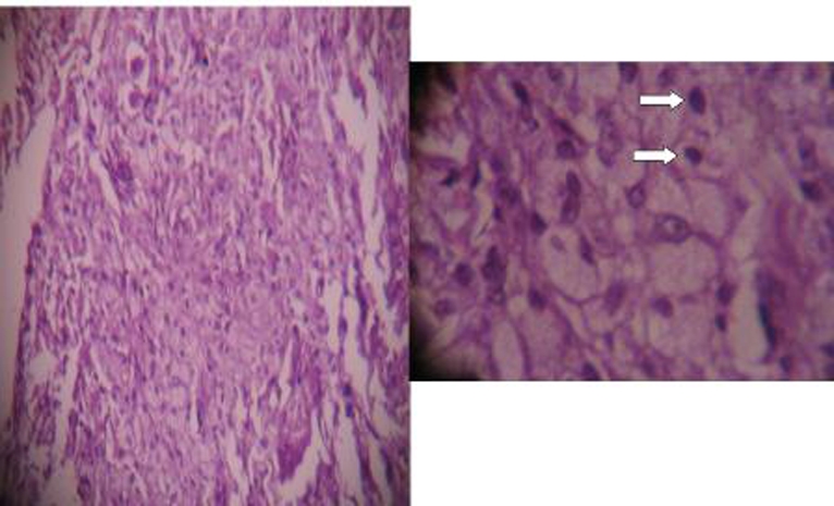
Figure 4 Histological features of tendinous xanthomatosis: showed proliferation of xanthomatous cells without atypia or mitoses. Abundant multinucleated Touton-like giant cells were observed with a mild interstitial inflammatory component. Arrows show foamy cells.
Author Disclosures: Rohan Arjun Bhojwani, Rajashree Khot
Please click here to visit the original article’s origin
Cerebrotendinous xanthomatosis: a rare genetic disorder – PubMed (nih.gov)